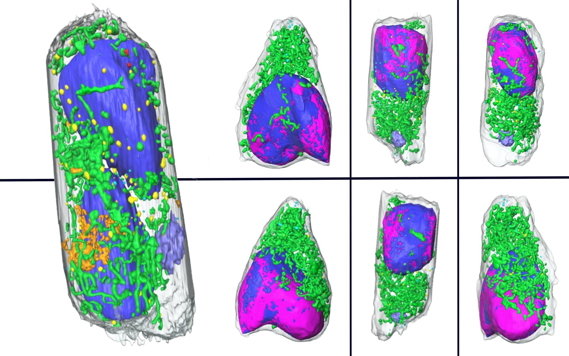
A major challenge in studying infectious diseases is identifying the changes cells undergo following invasion by pathogens. A highly sensitive technique developed in the early 2000s at the National Center for X-ray Tomography (NCXT), a facility based at Berkeley Lab’s Advanced Light Source (ALS), takes cellular imaging to the next level by sculpting three-dimensional scans of whole cells in mere minutes.
A team of researchers led by NCXT Director Carolyn Larabell, in collaboration with scientists at Heidelberg University in Germany, used this technique, called soft X-ray tomography (SXT), to quickly scan and analyze human lung cells infected with SARS-CoV-2. Prior to this technique, scientists would spend days or even weeks painstakingly preparing samples for electron microscopy to collect two-dimensional images of individual slices of the sample. These images then required further analysis to extrapolate three-dimensional structure.
SXT not only significantly shortens the time frame but provides more detail—increasing the chances of distinguishing subtle changes in the cell. “So, it really speeds up the process of examining cells, the consequences to infection, and the consequences of treating a patient with a drug that may or may not cure or prevent the disease,” Larabell, Head of the Cellular and Tissue Imaging Department in the Molecular Biophysics and Integrated Bioimaging Division, emphasized.
One intriguing structure identified by Jian-Hua Chen and Valentina Loconte, an affiliate senior scientist and postdoctoral researcher, respectively, in Larabell’s group, is a large membrane compartment in infected cells. Loconte suggested that it could be the cell’s way of recycling and removing viral replication machinery. The 3D tomographs produced by the SXT technique allowed for easier detection and tracking of this compartment compared to traditional electron microscopy methods.
Importantly, this approach also provides an avenue for others to conduct experiments on dangerous human pathogens, since the samples for SXT are chemically preserved. This is a key point given the significant risks and biosafety restrictions associated with working with live pathogens.
When SARS-CoV-2 emerged, scientists scrambled to understand how the novel virus impacts humans at a cellular level. The soft X-ray tomography technique not only opens the door for future studies in other infections but can also be used to investigate potential avenues for treatment.
Between the exquisite detail and the extremely fast turnaround, SXT could give researchers a significant advantage in the race against diseases like COVID-19.
Read more in the Berkeley Lab press release and BioScience News

