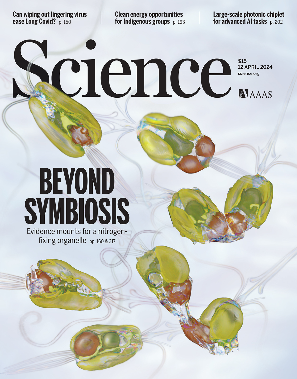
NCXT imaging technologies at your service
See primer for more detailsProtocol for full-rotation X-ray tomographySoft X-ray Tomography
Soft X-ray Tomography (SXT) is a method for imaging and quantifying the mesoscale structural organization inside a cell. Specimens are imaged after being cryo-fixed. They are, therefore, highly representative of a living cell. SXT can image cells up to 15 microns thick.
Super-resolution Cryolight Microscopy
Cryolight microscopy visualizes the location of labeled molecules inside a cell. Imaging at cryogenic temperature increases the working life of the fluorescent label, and prevents movement during imagine, as well as protecting the delicate cellular structures against damage.
Correlated Imaging
Specimens are imaged first with crygenic light microscopy and then by soft x-ray tomography. The two imaging data sets are then brought into alignment and overlaid. The resultant composite image builds a quantitative, high resolution structural model that contains the precise location of fluorescent-tagged molecules
Data Analysis
The final output of SXT is a 3D structural model of the specimen. This model has to be segmented into its components, such as the nucleus, mitochondria, and a host of other subcellular features. This has long been a time-consuming process. We are now automating this process by applying Machine Learning.
CASE STUDY
Pancreatic Beta Cell
A team of scientists — led by researchers at the Bridge Institute at the USC Michelson Center for Convergent Bioscience — have developed advanced imaging techniques to achieve the most detailed, multidimensional look ever inside pancreatic beta cells, the insulin factories of the body. They witnessed a choreography of reactions, some expected and others surprising, as the cell responded to changes.
Their work marks a new way to understand a disease that claims about 85,000 American lives annually. When applied to cells, these imaging breakthroughs are roughly akin to witnessing galaxies when astronomers gained advanced telescopes or germs when biologists got microscopes. Other techniques, often based on indirect measurement, tell part of the cell’s story, but the new direct and detailed observations provide a more complete picture.
Join us at the Microscopy&Microanalysis 2023 in Minneapolis, MN
Attend the Biological Soft X-ray Tomography symposium (B08) to learn more about the application of Soft X-ray tomography to Biology and learn about the the NCXT. Come and ask questions about the facility, or get expert advice on planning experiments, or analyzing date.
Recent research highlights
New model that simulates and quantifies the absorption of soft X-ray tomograms, allowing the interpretation of X-ray tomograms at the molecular level.
Autin, L., Barbaro, B., Jewett, A., Ekman, A., Verma, S., Olson, A., & Goodsell, D. (2022). Integrative structural modelling and visualisation of a cellular organelle. QRB Discovery, 3, E11. doi:10.1017/qrd.2022.10
Theoretical approach to evaluate the role of self-organized F-actin in motion generation and its impact at the molecular level would be reflected at the cellular scale
Hueschen, C.L., Zarko L.S., Chen, J.H., LeGros, M.A., Larabell, C.A., Boothroyd, J.C., Phillips, & R., Dunn, A.R. (2022). Emergent Actin Flows Explain Diverse Parasite Gliding Modes. bioRxiv, 495399; doi: 10.1101/2022.06.08.49539
Technical advancements in soft X-ray tomography
Ekman, A., Chen, J.-H., Vanslembrouck, B., Loconte, V., Larabell, C., Le Gros, M., & Weinhardt, V. (2022). Extending of imaging volume in soft x-ray tomography. Adv. Photonics Res. 2200142. doi:10.1002/adpr.202200142
Comprehensive and quantitative analysis of organelle interactions at the mesoscale
Loconte, V., Singla, J., Li, A., Chen, J.-H., Ekman, A., McDermott, G., Sali, A., Le Gros, M., White, K.L., & Larabell, C. A. (2022). Soft X-ray tomography to map and quantify organelle interactions at the mesoscale. Structure, 30, 1-12. doi:10.1016/j.str.2022.01.006
Cell Tomography Database
Research carried out at the NCXT is building a database of cell morphology, spanning in complexity from simple bacteria, through yeast and algea, to mammalian cells.
Image from Remesh et al. Nature Commuications, 11, 2905
Bacteria
Image from Walter et al. Mol Biol Cell, 30, 131-145. 10.1091/mbc.E18-04-0204
Yeast
Image from Aho et al, Viruses, 11, 10.3390/v11100935
Mammalian
Contact us
Funding
Use of the Advanced Light Source, an Office of Science User Facility operated for the U.S. Department of Energy (DOE) Office of Science by Lawrence Berkeley National Laboratory, was supported by the U.S. DOE under Contract No. DE-AC02-05CH11231.
This project was supported by grant (P30 GM138441) from the National Institute of General Medical Sciences of the National Institutes of Health.
The content is solely the responsibility of the authors and does not necessarily reflect the official views of NIGMS. the NIH, or the U.S. Department of Energy