Research

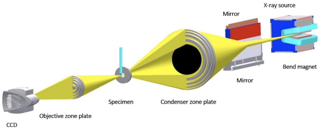
Super-resolution Cryolight Microscopy

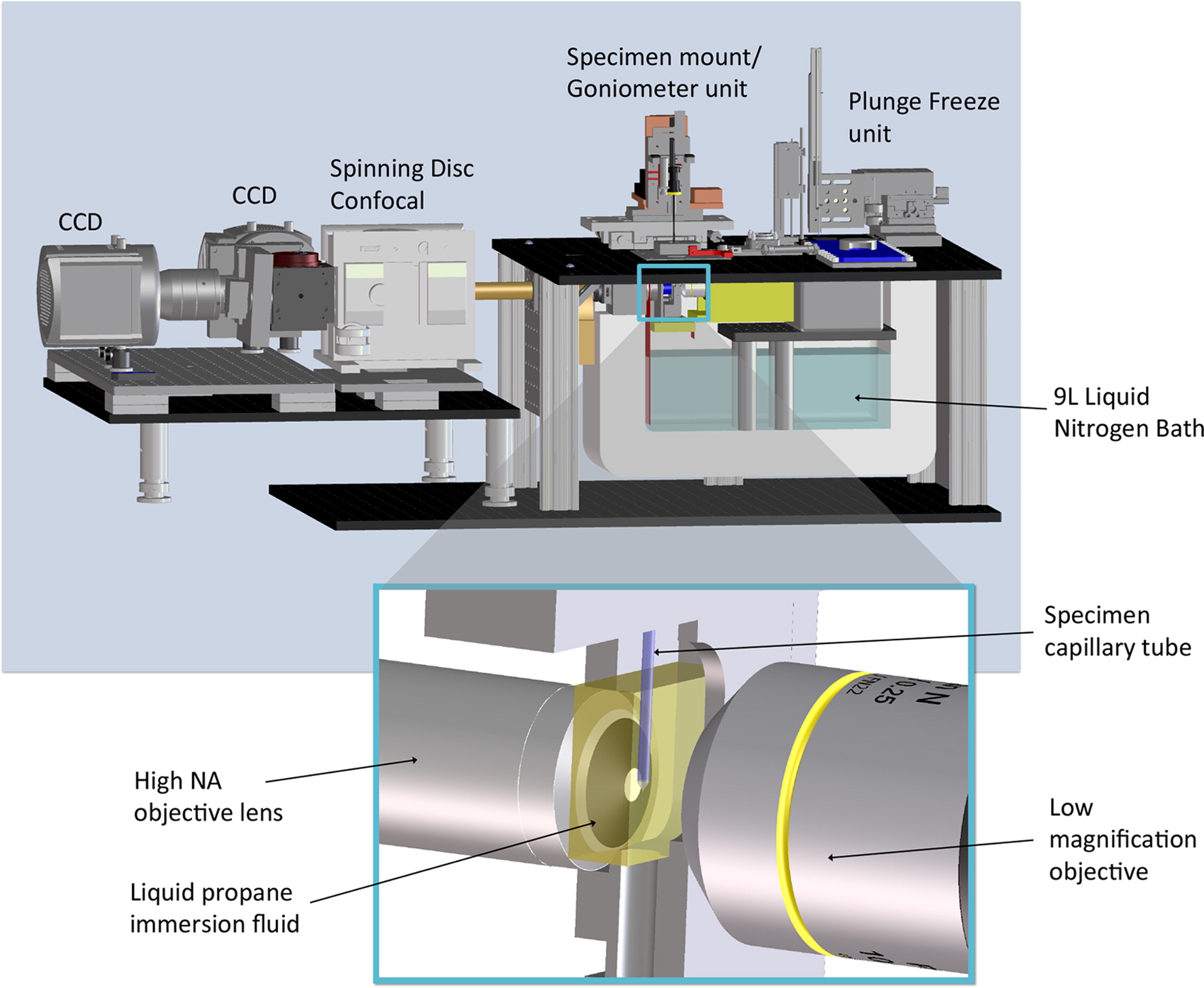
Fluorescence microscopy is a natural choice when combined with a modality designed for visualizing cell structure. The first-generation Fluorescence Cryo-Microscope (FCM) effectively addressed the challenge of imaging cryopreserved specimens in refractive index-matched fluids. This achievement was made possible by employing cryogenic immersion fluid, such as liquid propane or iso-pentane, instead of air, to enhance the [...]

Data Analysis

The final output of SXT is a 3D structural model of the specimen. This model has to be segmented into its components, such as the nucleus, mitochondria, and a host of other subcellular features. This has long been a time-consuming process. We are now automating this process by applying Machine Learning.

Correlated Imaging

Soft x-ray tomography can image an entire cell in its natural hydration state. Determining the location of specific molecular interactions requires that the molecular are labeled with a marker that is visible either in the soft x-ray microscope or in another modality and the data correlated. The latter is the preferred option since direct visualization [...]

Soft X-ray Tomography

Soft X-ray Tomography (SXT) is a non-invasive, 3D imaging technique that can measure volumes, surfaces, interfaces, membranes, and organelle connectivity within an intact cell. SXT data are collected on a transmission soft x-ray microscope, in the case of the NCXT, this is XM-2 at the Advanced Light Source, the world’s brightest source of x-rays. The [...]